Advance your Workflow - Abby
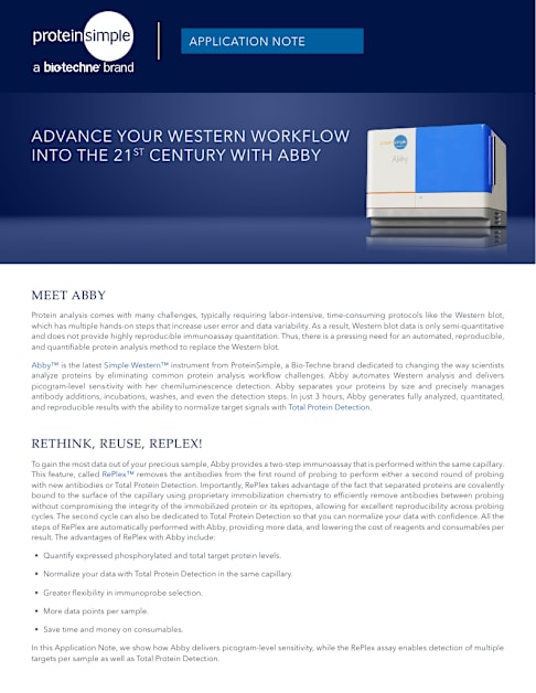
ADVANCE YOUR WESTERN WORKFLOW INTO THE 21ST CENTURY WITH ABBY
MEET ABBY Protein analysis comes with many challenges, typically requiring labor-intensive, time-consuming protocols like the Western blot, which has multiple hands-on steps that increase user error and data variability. As a result, Western blot data is only semi-quantitative and does not provide highly reproducible immunoassay quantitation. Thus, there is a pressing need for an automated, reproducible, and quantifiable protein analysis method to replace the Western blot.
Abby™ is the latest Simple Western™ instrument from ProteinSimple, a Bio-Techne brand dedicated to changing the way scientists analyze proteins by eliminating common protein analysis workflow challenges. Abby automates Western analysis and delivers picogram-level sensitivity with her chemiluminescence detection. Abby separates your proteins by size and precisely manages antibody additions, incubations, washes, and even the detection steps. In just 3 hours, Abby generates fully analyzed, quantitated, and reproducible results with the ability to normalize target signals with Total Protein Detection.
RETHINK, REUSE, REPLEX! To gain the most data out of your precious sample, Abby provides a two-step immunoassay that is performed within the same capillary. This feature, called RePlex™ removes the antibodies from the first round of probing to perform either a second round of probing with new antibodies or Total Protein Detection. Importantly, RePlex takes advantage of the fact that separated proteins are covalently bound to the surface of the capillary using proprietary immobilization chemistry to efficiently remove antibodies between probing without compromising the integrity of the immobilized protein or its epitopes, allowing for excellent reproducibility across probing cycles. The second cycle can also be dedicated to Total Protein Detection so that you can normalize your data with confidence. All the steps of RePlex are automatically performed with Abby, providing more data, and lowering the cost of reagents and consumables per result. The advantages of RePlex with Abby include:
• Quantify expressed phosphorylated and total target protein levels.
• Normalize your data with Total Protein Detection in the same capillary.
• Greater flexibility in immunoprobe selection.
• More data points per sample.
• Save time and money on consumables.
In this Application Note, we show how Abby delivers picogram-level sensitivity, while the RePlex assay enables detection of multiple targets per sample as well as Total Protein Detection.
APPLICATION NOTE
https://www.proteinsimple.com/abby.html?pdfSource=true_an_abby-simple-western https://www.proteinsimple.com/simple_western_overview.html?pdfSource=true_an_abby-simple-western https://www.proteinsimple.com/simple_western_overview.html?pdfSource=true_an_abby-simple-western https://shop.proteinsimple.com/total-protein-detection-module-for-wes-peggy-sue-or-sally-sue.html https://www.proteinsimple.com/jess_replex.html https://www.proteinsimple.com
2
NAME VENDOR PART NUMBER
12-230 kDa Abby, Jess or Wes Separation Module
ProteinSimple SM-W004
RePlex Module ProteinSimple RP001
Total Protein Detection Module ProteinSimple DM-TP01
Anti-Rabbit Detection Module for Abby, Jess, Wes, Peggy Sue or Sally Sue
ProteinSimple DM-001
Anti-Mouse Detection Module for Abby, Jess, Wes, Peggy Sue or Sally Sue
ProteinSimple DM-002
EGF Receptor Control Cell Extracts
Cell Signaling Technologies
5634
HeLa Lysate Santa Cruz Biotechnology
sc-24785
Recombinant DnaK Enzo Life Sciences ADI-SPP-630
MATERIALS AND METHODS The reagents used in this study are listed in TABLE 1 and the primary antibodies are listed in TABLE 2.
To determine the sensitivity of Abby, a 3-fold serial dilution series of recombinant DnaK was prepared in the presence of 0.1 mg/mL HeLa lysate, starting at 50000 pg/mL down to 69 pg/mL. Because only 3 µL of sample is required per well, this corresponds to 150 pg down to 0.21 pg, respectively.
All experiments were performed according to the default instructions as outlined in the 12-230 kDa Abby, Jess or Wes Separation, RePlex, and Total Protein Detection modules.
ABBY DELIVERS PICOGRAM SENSITIVITY With chemiluminescence detection, Abby provides highly sensitive detection of target proteins, even for proteins in complex samples like whole cell lysates. To determine the sensitivity of Abby’s chemiluminescence detection, we analyzed a serial dilution series of molecular chaperone DnaK spiked in HeLa lysate as described in the Materials and Methods in Probes 1 and 2 of the RePlex assay. As expected, a strong, clean signal corresponding to DnaK appeared with signal intensity that corresponded with sample concentration (FIGURE 1A). When peak area was plotted by DnaK concentration, a highly linear relationship was observed in both probing cycles, with R2 values of >0.997 (FIGURE 1B).
Importantly, the sensitivity and linearity of the assay were not compromised between Probes 1 and 2. Under these conditions, the limit of detection (LOD) was determined to be 2.6 pg in Probe 1 and 1.7 pg in Probe 2, respectively. Collectively, these results demonstrate that Abby delivers equivalent picogram-level sensitivity of detection in both Probe 1 and 2 of RePlex, allowing for the analysis of low abundance targets while conserving precious samples.
TABLE 1. Reagents used in this study.
TABLE 2. Primary antibodies used in this study. All antibodies were diluted in Antibody Diluent 2.
NAME VENDOR PART NUMBER DIL.
Anti-DnaK Novus Biologicals NBP1-97490 1:120
Anti-EGFR Novus Biologicals NBP2-34243 1:100
Anti-Phospho EGFR R&D Systems AF1095 1:200
Anti-GAPDH Novus Biologicals NB300-327 1:1000
Anti-HSP60 R&D Systems AF1800 1:100
Anti-Park7 Novus Biologicals NBP1-92715 1:1000
Anti-14-3-3 Novus Biologicals NB100-406 1:100
https://shop.proteinsimple.com/12-230-kda-wes-separation-module-8-x-25-capillary-cartridges.html?pdfSource=true_an_abby-simple-western https://shop.proteinsimple.com/replex-module-for-jess.html?pdfSource=true_an_abby-simple-western https://shop.proteinsimple.com/total-protein-detection-module-for-wes-peggy-sue-or-sally-sue.html?pdfSource=true_an_abby-simple-western https://shop.proteinsimple.com/anti-rabbit-detection-module-for-wes-peggy-sue-or-sally-sue.html?pdfSource=true_an_abby-simple-western https://shop.proteinsimple.com/anti-mouse-detection-module-for-wes-peggy-sue-or-sally-sue.html?pdfSource=true_an_abby-simple-western https://www.novusbio.com/products/hsp70-dnak-antibody-8e2-2_nbp1-97490?pdfSource=true_an_abby-simple-western https://www.novusbio.com/products/egfr-antibody-h9b4_nbp2-34243?pdfSource=true_an_abby-simple-western https://www.rndsystems.com/products/human-phospho-egfr-y1173-antibody_af1095?pdfSource=true_an_abby-simple-western https://www.novusbio.com/products/gapdh-antibody_nb300-327?pdfSource=true_an_abby-simple-western https://www.rndsystems.com/products/human-mouse-rat-hsp60-antibody_af1800?pdfSource=true_an_abby-simple-western https://www.novusbio.com/products/park7-dj-1-antibody-4h4_nbp1-92715?pdfSource=true_an_abby-simple-western https://www.novusbio.com/products/14-3-3-gamma-antibody-kc21_nb100-406?pdfSource=true_an_abby-simple-western
3
FIGURE 1. Abby chemiluminescent detection provides picogram sensitivity. (A) Lane view of DnaK detection in Probe 1 and 2 by the RePlex assay on Abby. (B) Linearity analysis of DnaK peak area by DnaK concentration from Probe 1 and Probe 2 of the RePlex assay on Abby.
FIGURE 2. Abby allows you to normalize your data with Total Protein Detection in the same capillary using RePlex. Raw and normalized signals of 14-3-3 are shown in varying concentrations of HeLa lysate. The signal of 14-3-3 was measured in Probe 1 and normalized to Total Protein Detection in Probe 2 of RePlex.
0
1000000
2000000
3000000
4000000
5000000
6000000
7000000
0.01 0.05 0.1 0.2
A ve
ra ge
P ea
k A
re a
Lysate concentration (mg/mL) Raw Normalized
expression data with confidence. To test this, HeLa lysates (0.01 – 0.2 mg/mL) were separated and probed with an anti-14-3-3 antibody in Probe 1 followed by the Total Protein Assay in Probe 2. While the peak area of unnormalized (raw) sample increased according to sample concentration, the protein normalization using RePlex shows comparable 14-3-3 signal across increasing concentrations of HeLa lysate (FIGURE 2). Because the expression of 14-3-3 is expected to remain stable regardless of the total protein concentration loaded, these results are consistent with accurate protein normalization.
RELIABLE PROTEIN NORMALIZATION WITH TOTAL PROTEIN DETECTION
To accurately quantify the expression of a target protein, it is necessary to normalize the signal of detection to a loading control. For this purpose, normalizing signal intensity to a housekeeping protein can be misleading, as the expression of housekeeping proteins can vary depending on the experimental conditions. Instead, normalizing protein expression to total protein content is a more reliable way to quantify the expression of a target protein.1 With RePlex, it is possible to normalize the expression of a target protein detected in Probe 1 with Total Protein Detection performed in Probe 2, so that you can normalize your protein
R² = 0.9979
R² = 0.9987
0
50000
100000
150000
200000
250000
300000
350000
400000
0 10 20 30 40 50 60
P ea
k A
re a
DnaK (pg)
Comparison of DnaK Peak Area in Probe 1 and Probe 2 of RePlex Assay
Probe 1 Probe 2
kDa 230-
180-
116-
66-
40-
12-
-DnaK
50 pg
16 .7
pg
5.6 pg
1.9 pg
50 pg
16 .7
pg
5.6 pg
1.9 pg
kDa 230-
180-
116-
66-
40-
12-
-DnaK
Probe 1 Probe 2
A B
4
FIGURE 3. RePlex gives you multiple measurements per sample, including detection of phosphorylated and total target protein levels. (A) Lane view of detection of EGFR and p-EGFR in Probe 1 and Probe 2, respectively, in EGF-treated and serum-starved (untreated) A431 lysates. (B) Fold change in expression of apparent p-EGFR signal and p-EGFR signal normalized to total EGFR in EGF-treated A431 cells. (C) Compass for Simple Western software automatically quantifies and normalizes protein expression. The Area column indicates raw peak area values for EGFR and p-EGFR and Corr. Area (Corrected Area) column indicates normalized peak area. In this example, EGFR in Probe 1 was normalized to EGFR signal in untreated A431 cells in capillary 5 (P1:5). Corrected Area for p-EGFR was then calculated based on the EGFR normalization. (D) Lane view of EGFR detection in Probe 1 followed by a secondary-only control in Probe 2 of RePlex to demonstrate antibody removal efficiency. (E) The anti-EGFR antibody removal efficiency between probing cycles of RePlex from EGF-treated and untreated A431 samples. The antibody removal efficiency was calculated as (Peak area Probe 1 – Peak area Probe 2)/Peak area Probe 1 x 100. Error bars represent standard deviations of the means.
DETECTION OF TOTAL AND PHOSPHO-PROTEIN PAIRS MADE EASY (FIGURE 3A).3 Next, we determined the fold change in apparent (unnormalized) p-EGFR and p-EGFR normalized to total EGFR. This analysis revealed that the apparent and normalized fold change were approximately 20 and 10, respectively (FIGURE 3B). Normalizing to total EGFR signal in both samples results in more accurate quantitation of the fold change in p-EGFR signal after EGF treatment. Gaining an accurate quantitative understanding of signaling networks as a whole is crucial and remains a daunting challenge, particularly with traditional Western blot.3 Thus, unraveling the signaling web in phosphoproteomics is just one application where RePlex can be leveraged as a robust assay.
kDa 230- 180-
116-
66-
40-
12-
-EGFR
EGF
kDa 230- 180-
116-
66-
40-
12-
pEGFR { EGFR
+ - + -EGF
0
5
10
15
20
25
Apparent Normalized to Total EGFR Signal
Fo ld
C ha
ng e
Fold Change in p-EGFR Signal in A431 Cells Treated with EGF
BA
C
ED
Probe 1: EGFR
Probe 2: p-EGFR
Probe 1: EGFR
Probe 2: 2°Ab only
+ - + -
0%
20%
40%
60%
80%
100%
120%
A431 + EGF A431 untreated
A nt
ib od
y R
em ov
al E
ffi ci
en cy
Anti-EGFR Antibody Removal
RePlex enables multiplex analysis of protein targets in the same capillary, even those with antibodies from the same host species or those that have overlapping molecular weights. For example, RePlex makes it easy to measure and compare total and phosphorylated levels of your target proteins. Here, we tested the ability to use EGFR and phospho-specific EGFR (p-EGFR) antibody pairs in lysates of EGF-treated and untreated A431 cells using RePlex. The conditions for this analysis were first optimized using the best practices outlined in the RePlex Method Development Guide.2 As expected, a strong EGFR signal was observed in EGF-treated and serum-starved (untreated) samples, while p-EGFR signal only appeared in the EGF-treated sample
https://www.proteinsimple.com/literature_download.html?docid=2156 https://www.proteinsimple.com/literature_download.html?docid=2156
5
To demonstrate that antibodies are efficiently removed between probing cycles on RePlex, we measured the removal of the anti- EGFR antibody from treated and untreated A431 by running a secondary-only control in Probe 2 of RePlex. Visual inspection showed that virtually no signal was detected in both samples when probed with the secondary antibody-only control in Probe 2 of RePlex (FIGURE 3D). Furthermore, quantitative analysis revealed that the antibody removal efficiency was >99% in both samples (FIGURE 3E). Thus, RePlex users can be confident that the antibodies used in Probe 1 will not interfere with analysis in Probe 2.
MULTIPLEX TO THE MAX Finally, we tested the ability of RePlex to multiplex targets in consecutive immunoassays. To do so, we measured 4 different targets (Park7, 14-3-3, GAPDH, and HSP60) in the same capillary. Two targets (Park7 and 14-3-3) were measured in Probe 1, and the other two targets (GAPDH and HSP60) were measured in Probe 2. Following RePlex analysis, all 4 targets were clearly separated and detected, and as expected, there was no interference of the antibody targets from Probe 1 in Probe 2 of RePlex (FIGURE 4). These data indicate that RePlex enables multiple targets to be probed within the same experiment, saving time and money while conserving precious samples.
Lean more | proteinsimple.com/abby.html Request pricing | proteinsimple.com/quote-request-simple-western-systems.html
FIGURE 4. RePlex was employed to detect 4 targets in the same sample and capillary. Graph vew (left) and lane view (right) of HSP60, GAPDH, 14-3-3, and Park7 detection performed across the two probing cycles of RePlex.
WESTERN BLOTS RE-IMAGINED Since its development approximately 50 years ago, the Western blot has hardly improved, burdening researchers with a long list of grievances. Today, researchers can leave the Western blot behind with Simple Western on Abby, which delivers picogram- level sensitivity, multiplex detection, and normalization with Total Protein Detection, in a single, fully automated platform. In just 3 hours (or 5 hours with RePlex), highly reproducible and quantifiable data are generated on up to 24 samples. The functionality of Abby is expanded with RePlex, to provide an additional round of immunoassay or Total Protein Detection, lowering the cost per result. Abby brings the Western blot into the 21st century.
REFERENCES 1. Transparency is the key to quality, A.J. Fosang and R.J. Colbran, The Journal of Biological Chemistry, 2015; 290:29692–4.
2. RePlex™ Method Development Guide, ProteinSimple, proteinsimple.com/literature_download.html?docid=2156
3. Cell signaling by receptor tyrosine kinases, M.A. Lemmon, J. Schlessinger, Cell, 2010; 141:1117-1134.
12 40 66 116 180 MW (kDa)
0
100,000
200,000
300,000
400,000
C he
m ilu
m in
es ce
nc e
P 1
0
100,000
200,000
300,000
400,000
500,000
C hem
ilum inescence P
2
Park7 14-3-3 GAPDH HSP60
kDa
180-
116-
66-
40-
12-
Pr ob
e 1
Pr ob
e 2
-HSP60
-GAPDH
-14-3-3
-Park7
Probe 1 Probe 2
https://www.proteinsimple.com/abby.html?pdfSource=true_an_abby-simple-western https://www.proteinsimple.com/quote-request-simple-western-systems.html https://www.proteinsimple.com/quote-request-simple-western-systems.html https://www.proteinsimple.com/quote-request-simple-western-systems.html https://pubmed.ncbi.nlm.nih.gov/26657753/ https://www.proteinsimple.com/literature_download.html?docid=2156 https://www.sciencedirect.com/science/article/pii/S0092867410006653
NOTES
6
NOTES
7
Template-ko
bio-techne.com Global info@bio-techne.com bio-techne.com/find-us/distributors TEL +1 612 379 2956 North America TEL 800 343 7475 Europe | Middle East | Africa TEL +44 (0)1235 529449 China info.cn@bio-techne.com TEL +86 (21) 52380373
For research use or manufacturing purposes only. Trademarks and registered trademarks are the property of their respective owners.
WHERE SCIENCE INTERSECTS INNOVATION™
AN_Abby-Simple-Western_STRY0110797
For more information visit or contact us at: Toll-free: 888 607 9692 Tel: 408 510 5500 info@proteinsimple.com proteinsimple.com
At ProteinSimple, we’re changing the way scientists analyze proteins. Our innovative product portfolio helps researchers reveal new insight into proteins, advancing their understanding of protein function. We enable cutting-edge research to uncover the role of proteins in disease and provide novel approaches to develop and analyze protein-based therapeutics. We empower you to make your next discovery by eliminating common protein analysis workflow challenges.
https://www.bio-techne.com https://www.rndsystems.com https://www.novusbio.com https://www.tocris.com https://www.proteinsimple.com https://acdbio.com https://www.exosomedx.com https://www.bio-techne.com