Multi-Antigen Serology Assay using Simple Western
A MULTI-ANTIGEN SEROLOGY ASSAY FOR COVID-19 USING SIMPLE WESTERN
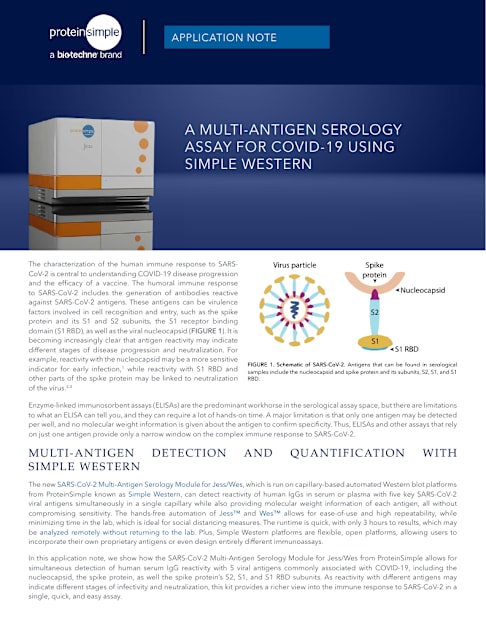
The characterization of the human immune response to SARS- CoV-2 is central to understanding COVID-19 disease progression and the efficacy of a vaccine. The humoral immune response to SARS-CoV-2 includes the generation of antibodies reactive against SARS-CoV-2 antigens. These antigens can be virulence factors involved in cell recognition and entry, such as the spike protein and its S1 and S2 subunits, the S1 receptor binding domain (S1 RBD), as well as the viral nucleocapsid (FIGURE 1). It is becoming increasingly clear that antigen reactivity may indicate different stages of disease progression and neutralization. For example, reactivity with the nucleocapsid may be a more sensitive indicator for early infection,1 while reactivity with S1 RBD and other parts of the spike protein may be linked to neutralization of the virus.2,3
Enzyme-linked immunosorbent assays (ELISAs) are the predominant workhorse in the serological assay space, but there are limitations to what an ELISA can tell you, and they can require a lot of hands-on time. A major limitation is that only one antigen may be detected per well, and no molecular weight information is given about the antigen to confirm specificity. Thus, ELISAs and other assays that rely on just one antigen provide only a narrow window on the complex immune response to SARS-CoV-2.
MULTI-ANTIGEN DETECTION AND QUANTIFICATION WITH SIMPLE WESTERN The new SARS-CoV-2 Multi-Antigen Serology Module for Jess/Wes, which is run on capillary-based automated Western blot platforms from ProteinSimple known as Simple Western, can detect reactivity of human IgGs in serum or plasma with five key SARS-CoV-2 viral antigens simultaneously in a single capillary while also providing molecular weight information of each antigen, all without compromising sensitivity. The hands-free automation of Jess™ and Wes™ allows for ease-of-use and high repeatability, while minimizing time in the lab, which is ideal for social distancing measures. The runtime is quick, with only 3 hours to results, which may be analyzed remotely without returning to the lab. Plus, Simple Western platforms are flexible, open platforms, allowing users to incorporate their own proprietary antigens or even design entirely different immunoassays.
In this application note, we show how the SARS-CoV-2 Multi-Antigen Serology Module for Jess/Wes from ProteinSimple allows for simultaneous detection of human serum IgG reactivity with 5 viral antigens commonly associated with COVID-19, including the nucleocapsid, the spike protein, as well the spike protein’s S2, S1, and S1 RBD subunits. As reactivity with different antigens may indicate different stages of infectivity and neutralization, this kit provides a richer view into the immune response to SARS-CoV-2 in a single, quick, and easy assay.
Virus particle
S1
S2
Spike protein
S1 RBD
Nucleocapsid
FIGURE 1. Schematic of SARS-CoV-2. Antigens that can be found in serological samples include the nucleocapsid and spike protein and its subunits, S2, S1, and S1 RBD.
APPLICATION NOTE
https://www.proteinsimple.com/sars-cov-2-multi-antigen-serology-module.html https://www.proteinsimple.com/simple_western_overview.html https://www.proteinsimple.com/jess.html https://www.proteinsimple.com/wes.html https://www.proteinsimple.com/literature_download.html?docid=2123 https://www.proteinsimple.com
2
HOW THE SARS-CoV-2 MULTI-ANTIGEN SEROLOGY MODULE WORKS The SARS-CoV-2 Multi-Antigen Serology Module is compatible with any Jess or Wes instrument and is used in combination with the 12-230 kDa Jess/Wes Separation Module (PN SM-W004). This module enables detection of human IgG antibodies in human serum or plasma reactive against multiple SARS-CoV-2 antigens with characterized IgG class specificity. A workflow of how the SARS-CoV-2 Multi-Antigen Serology Module compares to a standard Simple Western assay is shown in FIGURE 2.
FIGURE 2. How the SARS-CoV-2 Multi-Antigen Serology Module works compared to a standard Simple Western assay. First, the antigens are separated and immobilized to the capillary wall. Then, serum or plasma is used in place of the primary antibody solution, followed by immunodetection with an anti-IgG HRP conjugate and chemiluminescent detection.
MATERIALS
The following components are included in the SARS-CoV-2 Multi- Antigen Serology Module for Jess/Wes (SA-001):
SARS-CoV-2 MULTI-ANTIGEN LADDER KIT (SA-001-1)
• SARS-CoV-2 S1 Subunit RBD
• SARS-CoV-2 Nucleocapsid Protein
• SARS-CoV-2 Spike S1 Subunit Protein
• SARS-CoV-2 Spike S2 Subunit Protein
• SARS-CoV-2 Spike Protein
• Human anti-His primary antibody
ANTI-HUMAN IgG DETECTION MODULE (DM-005)
• Anti-Human IgG Secondary HRP Antibody
• Luminol-S, Peroxide
• Antibody Diluent 2
• Streptavidin-HRP
SEROLOGY MODULE DILUENTS (SA-001-2)
• Serum Diluent
• Reconstitution Reagent 2
METHODS
SERUM SAMPLES TREATMENT
Using a serum separator tube (SST), samples were allowed to clot for 30 minutes at room temperature before centrifugation for 15 minutes at 1000 x g.
Serum was removed and heat inactivated immediately (below), or aliquoted and stored at ≤ -20 °C for later heat inactivation to avoid repeated freeze-thaw cycles.
HEAT INACTIVATION PROTOCOL
For fresh serum samples, heat inactivation was performed at 56 °C for 1 hour. Frozen serum samples were allowed to thaw before heat inactivation at 56 °C for 1 hour. Samples were aliquoted and stored at ≤ -20 °C.
SAMPLE DILUTION
To create one well of sample at a 1:10 dilution, 2 µl of serum was diluted with 18 µL of serum diluent and gently mixed. For additional wells, volumes were adjusted accordingly.
Low MWHigh MW
Stacking Matrix Separation Matrix
Load Matrix
Load Sample
Separate
Immobilize
Immunoprobe with Primary Antibody
Quantitate
Load Matrix
Load Antigen
Separate
Immobilize
Immunoprobe with Serum
Quantitate
Target Protein Primary Ab
HRP-labeled Secondary Ab
12 40 66 90 116 180
MW (kDa)
Size Separation Standard
Assay Serum Assay
Protein
C h
em ilu
m in
es ce
n ce
3
PUTTING THE SARS-COV-2 MULTI-ANTIGEN SEROLOGY MODULE TO THE TEST
FIGURE 3. Detection of all five antigens in a single capillary. The electropherogram resulting from this analysis is shown on the left and the lane view is shown on right. Each antigen was detected with a humanized anti-His primary antibody and anti-human IgG HRP conjguate supplied in the SARS-CoV-2 Multi-Antigen Serology Module.
12 40 66 116 180 230 MW (kDa)
0
100,000
200,000
300,000
400,000
C he
m ilu
m in
es ce
nc e
S1 RBD Nucleocapsid
S2 Subunit S1 Subunit Spike
-Spike
-S1 Subunit
-S2 Subunit
-Nucleocapsid
-S1 RBD
kDa 230-
180-
116-
66-
40-
12-
Bi oti
n L ad
de r
SA RS
-C oV
-2 La
dd er
TABLE 1. Apparent molecular weight of each antigen.
ANTIGEN MW (kDA)
Spike 170
S1 Subunit 98
S2 Subunit 69
Nucleocapsid 57
S1 RBD 47
Due to differences in molecular weight (MW), all five antigens of the SARS-CoV-2 Multi-Antigen Serology Module can be resolved and detected in a single capillary (TABLE 1). To demonstrate this, we analyzed all five antigens simultaneously on Jess. Each antigen has a His-tag that was exploited for detection using the human anti-His primary antibody and anti-human IgG HRP-conjugated secondary antibody supplied in the module (FIGURE 3). This analysis showed clearly defined peaks for each of the 5 antigens, indicating that all 5 antigens can be resolved and identified simultaneously on Jess.
4
FIGURE 4. PCR positive serum samples reacting with antigens. All serum samples were tested at 1:10 dilution in Serum Diluent. The overlaid electropherograms resulting from this analysis are shown on the left and the lane view is shown on the right.
To test reactivity of serum samples against these antigens, we analyzed serum samples that were confirmed positive for COVID-19 by polymerase chain reaction (PCR). In this analysis, serum samples are used as the primary antibody, and the secondary anti-human IgG HRP conjugate supplied in the module was used for chemiluminescence detection, as was described in FIGURE 2. Two PCR-negative serum samples were included as negative controls. This analysis showed a high degree of reactivity of the PCR positive serum samples with the antigens included in the kit (FIGURE 4). As expected, little to no reactivity occurred with the PCR negative serum samples. When relative peak areas were quantified, this analysis revealed a high degree
12 40 66 116 180 230 MW (kDa)
0
100,000
200,000
300,000
400,000
500,000
600,000
700,000
C he
m ilu
m in
es ce
nc e
S1 RBD Nucleocapsid
S2 Subunit
S1 Subunit Spike
PCR Pos 1
PCR Pos 2
PCR Pos 3
PCR Pos 4
PCR Neg 1
PCR Neg 2
-Spike
-S1 Subunit
-S2 Subunit
-Nucleocapsid
-S1 RBD
kDa 230-
180-
116-
66-
40-
12-
PC R
Po s 1
PC R
Po s 2
PC R
Po s 3
PC R
Po s 4
PC R
Ne g 1
PC R
Ne g 2
SARS-CoV-2 Antigen Detection for PCR Positive Human Sera
0
20
40
60
80
100
120
PCR Pos 1 PCR Pos 2 PCR Pos 3 PCR Pos 4
% P
ea k
A re
a
Serum Sample
Spike S1 Subunit S2 Subunit Nucleocapsid S1 RBD
FIGURE 5. Quantification of percent peak area for SARS-CoV-2 antigens detected by PCR positive human sera.
of variability in IgG reactivity with each of the 5 antigens among the human serum samples (FIGURE 5). For example, PCR Pos 4 had high reactivity (68% of total peak area) with the nucleocapsid, while PCR Pos 1, 2, and 3 had much lower reactivity (7, 3, and 34%, respectively) with the nucleocapsid, but increased reactivity with the spike protein. This is critical information as it was shown that detection of the nucleocapsid is sensitive to early infection,1
while antibodies that target epitopes on the spike protein may be linked to neutralization.2-3 Overall, these data provided a much deeper characterization of the humoral response to COVID-19 than is possible with single-antigen ELISA kits.
5
FIGURE 7. The linear range of detection as determined by a serial dilution series of PCR positive serum. (A) Overlaid electropherograms of serum dilutions 1:40 - 1:8640. (B) Overlaid electropherograms of serum dilutions 1:8460 - 1:138240. (C) Lane view of serum dilutions 1:40 - 1:138240. (D) The average peak area of two duplicates was calculated for each serum dilution, resulting in linear relationships between signal intensity and dilution factor for all five antigens. Error bars represent the standard deviations of the means.
Next, we determined how the SARS-CoV-2 Multi-Antigen Serology Module compares to an ELISA by using the COVID- SeroIndex™ Kantaro Quantitative SARS-CoV-2 IgG Antibody RUO ELISA Kit (R&D Systems, PN DSR200). We tested samples identified as positive and negative based on the RBD and Spike ELISA orthogonal testing method and calculated the positive percent agreement (PPA) and negative percent agreement (NPA)
12 40 66 116 180 230 MW (kDa)
0
100,000
200,000
300,000
400,000
500,000
600,000
700,000
C he
m ilu
m in
es ce
nc e
S1 RBD
Nucleocapsid
S2 Subunit
S1 Subunit Spike
12 40 66 116 180 230 MW (kDa)
0
2,000
4,000
6,000
8,000
10,000
12,000
14,000
16,000
C he
m ilu
m in
es ce
nc e S1 RBD
Nucleocapsid
S2 Subunit S1 Subunit
Spike
R² = 0.9917
R² = 0.9916
R² = 0.9989
R² = 0.9821
R² = 0.9928
100
1000
10000
100000
1000000
10000000
100000000
10 100 1000 10000 100000 1000000
A ve
ra ge
P ea
k A
re a
Serum Dilution
Linearity of SARS-CoV-2 Antibodies in Human Serum
S1 RBD
Nucleocapsid
S2 Subunit
S1 Subunit
Spike
-Spike
-S1 Subunit
-S2 Subunit
-Nucleocapsid
-S1 RBD
kDa
230-
180-
116-
66-
40-
1:4 0
1:1 20
1:3 60
1:1 08
0
1:2 16
0
1:4 32
0
1:8 64
0
1:1 72
80
1:3 45
60
1:6 91
20
1:1 38
24 0
1:40 1:120 1:360 1:1080 1:2160 1:4320 1:8640
1:8640 1:17280 1:34560 1:69120 1:138240
A B
C D
Serum dilution: Serum dilution:
using the Kantaro ELISA as the reference method. In this study, samples from 137 individuals were tested using the SARS-CoV-2 Multi-Antigen Serology Module, with 62 individuals previously identified as positive and 75 previously identified as negative. A total of 166 tests were performed using these samples, with 66 tests from the positive samples and 100 tests from the negative samples, as some samples were tested more than once on Jess. For this sample set, it was determined that the appropriate cut-off value for identifying negative and positive samples when using the SARS-CoV-2 Multi-Antigen Serology Module was 1,200,000 total peak area, which combines the peak area for all 5 antigens as measured by Compass for Simple Western software. This cut- off was determined using JMP statistical software and generating a Receiver Operating Characteristic (ROC) curve for the collected data. Using the appropriate cut-off, we showed that there was 97% PPA and 99% NPA between the SARS-CoV-2 Multi-Antigen Serology Module and the Kantaro ELISA (TABLE 2).
Finally, we determined the linear range of the SARS-CoV-2 Multi- Antigen Serology Module. To test this, we prepared a dilution series of a PCR positive serum sample from 1:40 to 1:138240 in duplicate and analyzed this with the SARS-CoV-2 Multi- Antigen Serology Module. As expected, signal decreased with decreasing serum sample concentration (FIGURE 7A-C), and the assay showed great linearity, with R2 ≥ 0.99 for four of the five antigens across the 3.5 log titration series, and R² ≥ 98% for the fifth antigen (FIGURE 7D).
TABLE 2. Positive percent agreement (PPA) and negative percent agreement (NPA) of the SARS-CoV-2 Multi-Antigen Serology Module compared to the Kantaro COVID-SeroIndex ™ RUO ELISA. In this study, 137 individuals identified as positive (62) or negative (75) based on the Kantaro RBD and Spike ELISA were analyzed using the SARS-CoV-2 Multi-Antigen Serology Module, in which some samples were run more than once.
BENCHMARKING AGAINST THE KANTARO COVID-SEROINDEX™ RUO ELISA
Kantaro COVID-SeroIndex™ RUO ELISA
Positive Negative
SARS-CoV-2 Multi-Antigen
Serology Module for Jess/Wes
Positive 64 1
Negative 2 99
Total 66 100
PPA 97%
NPA 99%
https://www.rndsystems.com/products/kantaro-ruo-sars-cov-2-igg-elisa-100%2525-seroprevalence-_dsr200 https://www.rndsystems.com/products/kantaro-ruo-sars-cov-2-igg-elisa-100%2525-seroprevalence-_dsr200 https://www.rndsystems.com/products/kantaro-ruo-sars-cov-2-igg-elisa-100%2525-seroprevalence-_dsr200
6
FIGURE 8. Peak area percent change to previous dilution in the linearity analysis of the serial dilution series. First, peak areas were normalized based on dilution. For example, for the 1:360 dilution sample, each antigen’s peak area was multiplied by 360. Then, the ratio of an antigen’s normalized peak area for sequential dilutions was calculated (e.g., the 1:360 dilution was divided by the 1:120 dilution), and this ratio was plotted for each serum dilution. The dotted black lines indicate ± 20% of the target ratio of 100%, which is considered an acceptable range in related ELISAs.
0%
20%
40%
60%
80%
100%
120%
140%
160%
180%
200%
220%
P ea
k A
re a
P er
ce nt
C ha
ng e
to P
re vi
ou s
D ilu
tio n
Serum Dilution
Dilution Linearity for SARS-CoV-2 Reactive Human Serum
S1 RBD
Nucleocapsid
S2 Subunit
S1 Subunit
Spike
1:1 20
1:3 60
1:1 08
0
1:2 16
0
1:4 32
0
1:8 64
0
1:1 72
80
1:3 45
60
1:6 91
20
1:1 38
24 0
TABLE 3. CVs resulting from duplicate measurements of the serum dilution series. At a 1:138240 dilution, the S1 RBD and S2 Subunit antigens are near the detection limit and therefore have high CVs. CVs below 20% are considered acceptable in related ELISAs.
SERUM DILUTION S1 RBD NUCLEOCAPSID S2 SUBUNIT S1 SUBUNIT SPIKE
1:40 5% 3% 3% 4% 5%
1:120 5% 4% 6% 6% 5%
1:360 3% 2% 3% 3% 4%
1:1080 4% 3% 3% 4% 3%
1:2160 4% 2% 4% 4% 6%
1:4320 3% 1% 2% 4% 4%
1:8640 3% 2% 3% 4% 7%
1:17280 7% 1% 2% 8% 8%
1:34560 11% 0% 3% 2% 3%
1:69120 14% 1% 11% 5% 1%
1:138240 76% 4% 46% 4% 7%
For a linear series, the ratio of an antigen’s normalized peak area for sequential dilutions (e.g., 1:360 dilution divided by 1:120) should be close to 100%, and a range of 80-120% is typically acceptable in related ELISAs. Based on these criteria, dilutions between 1:1080 and 1:69120 are considered to be in the linear
range for all 5 antigens for this serum sample (FIGURE 8). In addition, CVs were less than 15% for serum dilutions where peaks were above the limit of detection, or ≥1:69120 (TABLE 3), which demonstrates excellent reproducibility among duplicate measurements in the assay.
7
MORE INSIGHT INTO THE COVID-19 IMMUNE RESPONSE WITH LESS SERUM As COVID-19 progresses in the body, several different antigens may be present that illicit a humoral immune response, and the emergence of antibodies that target these antigens may indicate different stages of disease progression and virus neutralization.1–3
Therefore, ELISA microplate assays coated with only one antigen can capture only a very limited snapshot of a much broader immune response. With the SARS-CoV-2 Multi-Antigen Serology Module for Jess/Wes, up to 5 different antigens that commonly appear in COVID-19 can be analyzed for IgG reactivity in human serum or plasma samples simultaneously. This enables a much deeper analysis of the humoral response and it reveals variability among serum reactivity that cannot be captured by commercial microplate ELISA kits. Furthermore, the assay developed here uses significantly less serum than commercial ELISA kits. For instance, many ELISAs typically require 10 µL of serum, but the SARS-CoV-2 Multi-Antigen Serology Module can use as little as 1 µL of serum based on the minimum recommended dilution of 1:10. However, much lower titers can be detected and still be within the linear range of detection, as we’ve demonstrated in this application note. Taken together, in a single 3-hour automated runtime, reactivity with 5 different antigens on up to 23 samples can be analyzed using a tiny amount of serum, enabling rich and rapid characterization of the humoral response to COVID-19.
Order serology assay kit
REFERENCES 1. Sensitivity in detection of antibodies to nucleocapsid and spike proteins of severe acute respiratory syndrome coronavirus 2 in patients with coronavirus disease 2019, P Burbelo, F Riedo, C Morishima, S Rawlings, D Smith, S Das, J Strich, D Chertow, R Davey and J Cohen, The Journal of Infection Diseases, 2020; 222:206-213.
2. Isolation of potent SARS-CoV-2 neutralizing antibodies and protection from disease in a small animal model, T Rogers, F Zhao, D Huang, N Beutler, A Burns, W He, O Limbo, C Smith, G Song, J Woehl, L Yang, R Abbott, S Callaghan, E Garcia, J Hurtado, et al., Science, 2020; 369(6506):956–963.
3. A panel of human neutralizing mAbs targeting SARS-CoV-2 spike at multiple epitopes, T Noy-Porat, E Makdasi, R Alcalay, A Mechaly, Y Levy, A Bercovich-Kinori, A Zauberman, H Tamir, Y Yahalom-Ronen, M Israeli, E Epstein, H Achdout, S Melamed, T Chitlaru, S Weiss, et al., Nature Communications, 2020; 11(1):1–7.
Request pricing
https://shop.proteinsimple.com/sars-cov-2-multi-antigen-serology-module-for-jess-wes.html https://academic.oup.com/jid/article/222/2/206/5840542 https://academic.oup.com/jid/article/222/2/206/5840542 https://academic.oup.com/jid/article/222/2/206/5840542 https://www.ncbi.nlm.nih.gov/pmc/articles/PMC7299280/ https://www.nature.com/articles/s41467-020-18159-4 https://www.proteinsimple.com/quote-request-simple-western-systems.html
Template-ko
bio-techne.com Global info@bio-techne.com bio-techne.com/find-us/distributors TEL +1 612 379 2956 North America TEL 800 343 7475 Europe | Middle East | Africa TEL +44 (0)1235 529449 China info.cn@bio-techne.com TEL +86 (21) 52380373
For research use or manufacturing purposes only. Trademarks and registered trademarks are the property of their respective owners.
WHERE SCIENCE INTERSECTS INNOVATION™
AN_COVID-19-Immune-Response-Simple Western_STRY0084571
For more information visit or contact us at: Toll-free: 888 607 9692 Tel: 408 510 5500 info@proteinsimple.com proteinsimple.com
At ProteinSimple, we’re changing the way scientists analyze proteins. Our innovative product portfolio helps researchers reveal new insight into proteins, advancing their understanding of protein function. We enable cutting-edge research to uncover the role of proteins in disease and provide novel approaches to develop and analyze protein-based therapeutics. We empower you to make your next discovery by eliminating common protein analysis workflow challenges.
https://www.bio-techne.com https://www.rndsystems.com https://www.novusbio.com https://www.tocris.com https://www.proteinsimple.com https://acdbio.com https://www.exosomedx.com https://www.bio-techne.com mailto:info%40proteinsimple.com%0D?subject= https://www.proteinsimple.com/